Why and when it is important to use imaging systems in pre-clinical trials
The whole purpose of pre-clinical trials is to test a medical intervention for its safety and efficacy before clinical testing on humans. Using imaging systems before, during and after a pre-clinical trial provides essential data on the effects of the device, procedure or drug being tested.
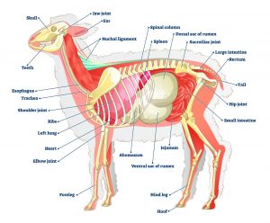
While it is easy to understand the value of imaging during and after the trial, some might question the need for using imagery on an animal before beginning the trial. In addition to giving a precise view of the organ or system involved, deciding to use imaging prior to the trial ensures that the chosen animal is indeed anatomically suited for the trial:
- to decide exactly where to place the device;
- to pinpoint the procedure area;
- to determine the best method of accessing the particular organ or system.
Additionally, collecting imaging prior to beginning the trial allows for a definitive before and after comparison of the organs and/or systems involved in the trial.
What are the main types of imaging systems used in preclinical trials
Ultrasound
An ultrasound or sonography is a device that uses sound waves to illustrate the internal organs, assess their function, as well as show blood flow and are particularly useful for identifying the site of various procedures. These can be used to obtain a baseline (B.L.) for implantations (i.e., valve replacement).
Ultrasound machines can be equipped for either two-dimensional or three-dimensional displays. At Lahav CRO, we have both to allow for either option.
Ultrasound probes
Ultrasound machines are used together with probes, each probe allowing for a different use and view of the organ or procedure.
The TTE (Trans Thoracic Echo) probe is for external use and not used during procedures. It is not used extensively, since the echogenic view is limited in large animal models e.g. ovine and ewes (e.g, no apical views).
The TEE (trans esophageal echo) probe is used internally. It can be inserted into the esophagus or directly into the heart via an opening in the chest. Due to its ease of use, the TEE probe is widely used during surgery, to capture an exact picture of the organ, device and/or procedure.
The ICE (Intra Cardiac Echo) probe can be inserted into either a vein or artery, giving a somewhat limited image. However, there are procedures (i.e. transeptal) that the ICE probe is the probe of choice. In these cases, the ICE probe enables the capturing of an image without the need for an additional chest surgery.
It is worth noting Lahav’s recent acquisition of the EPIQ CVx, a dedicated cardiac ultrasound solution which brings significant advancements in functionality. The CVx ultrasound, can produce diagnostic images for cardiac, abdominal, musculoskeletal, urology, transvaginal, and small organs, as well as a 3D anatomical structure of a fetus or organ. With this ultrasound machine, adjustments can be made to light and shadow, while viewing 3D images.
X-Ray
While X-ray machines have been around since 1895,1 their usefulness today cannot be denied. There have been many changes and improvements since they first came into use. Since X-Rays only provide a two-dimensional image, it is often used together with the ultrasound to provide a more complete view. The X-Ray machine is useful for monitoring implanted devices, to ensure that they have not migrated away from the intended implant location. Additionally, contrast material may be used to better identify the targeted anatomy and organs.
Our facility X-Ray machine is The Philips Zenition 70 with a C-arm which makes it easy and comfortable to use in surgeries and interventions.
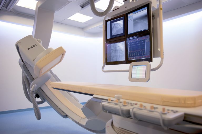
The Catheterization Lab
The catheterization unit is a stationary system with complex functions that allow for higher quality images of the circulatory system, as well as for catheterized based transplants. Our catheterization unit produces images (50-60 KW) that are 3-4 times higher quality than those obtained with regular fluoroscopy. This more advanced system answers the needs that arise when working with the larger animals.
We deploy a Philips ALLURA FD20, with XperCT settings, providing our clients an additional layer of imaging to streamline procedures. The XperCT provides high-quality imaging in the interventional suite, enabling clinicians to assess soft tissue, bone structure and stent deployment – before, during and after interventional procedures.
We are happy to offer an upgraded digital processing system, which has reduced radiation exposure, making the lab safer for those working in the lab.
Computed Tomography Scan (CT)
Our post operative suite is equipped with a state-of-the-art Philips Brilliance 64 CT scanner. This advanced imaging technology enables precise 3D anatomical modeling, essential for detailed planning in device and procedural trials. With the ability to capture high-resolution images of critical areas such as the heart, brain, and lungs in approximately five seconds, the 64-slice scanner covers about 40 millimeters per pass, ensuring comprehensive and accurate imaging.
Post-procedure CT scans are pivotal for verifying blood vessel patency and confirming the location and position of devices. The high-resolution capabilities of the Philips Brilliance 64 CT scanner support meticulous monitoring and assessment, enhancing the reliability and outcomes of clinical trials. The images provided by CT’s allow for the building of anatomy (3D) to the specific measurement requirements for precise planning for the device and procedural trials. Post procedure CT enables to check blood vessels patency and device location and position.
Imaging at Lahav CRO
The use of imagery in preclinical trials has been well documented, with similar conclusions. Preclinical imaging technologies allow for the non-invasive evaluation of tissue structure, and function in animal subjects. They bolster preclinical research by enabling real-time monitoring of pharmacological, device and procedural effects.2,3,4
Indeed, Wong et. al. goes so far as to say that ‘imagery plays a fundamental role’ as it relates to preclinical trials for drug development.5
Lahav CRO research center is equipped with all of the imaging systems described above. While having a wide range of imaging methods to choose from may be confusing initially – Our team is made up of biomedical research experts, with a broad range of expertise: primarily cardiovascular, Gastrointestinal, renal and urinary tract disorders, ophthalmology, dental, and orthopedic. This means that in addition to having the facilities needed to conduct your chosen pre-clinical trial at the highest level, hawse also have the experts to advise you and your team on the best methods to receive the most comprehensive results for a successful pre-clinical trial.
- Tubiana, Maurice. “Wilhelm Conrad Röntgen and the discovery of X-rays.” Bulletin de l’Academie nationale de medecine 180.1 (1996): 97-108.
- Hunter, Philip. “Illuminating human disease: The potential of in vivo imaging for preclinical research and diagnostics.” EMBO reports 20.10 (2019): e49195.
- Matthews, Paul M., et al. “Technologies: preclinical imaging for drug development.” Drug Discovery Today: Technologies 10.3 (2013): e343-e350.
- Bilgen, Mehmet. “Feasibility and merits of performing preclinical imaging on clinical radiology and nuclear medicine systems.” International Journal of Molecular Imaging 2013 (2013).
- Wong, Dean F. “Imaging in drug discovery, preclinical, and early clinical development.” The Journal of Nuclear Medicine 49.6 (2008): 26N.