Sheep as an animal model for orthopedic studies – The suitability of sheep for orthopedic models.
In our previous blog, we discussed how to choose a large animal for preclinical studies and focused on Sheep models. Here we will delve in deeper and discuss ‘why’ and ‘what’ makes sheep models suitable for preclinical studies, especially in the field of orthopedics.
Comparative anatomy – spine
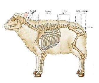
While it might not seem obvious, the anatomical bone structure of sheep is indeed similar to human bone structure, especially the spine. Given that sheep do not stand in an upright position (vertical posture), the visual below is intended to provide insight.

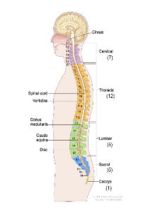
In the above diagram, the parallel can be drawn between the spines of the sheep and humans. Below, the spinal vertebra.
| Number in Sheep | Number in Humans | Vertebra |
| 7 | 7 | Cervical |
| 13-14 | 12 | Thoracic |
| 6-7 | 5 | Lumbar |
| 4 | 5 | Sacral |
| 7 | 1 | Coccyx |
The sheep spine is a valuable model for experiments related to the gross structure of the thoracic, lumbar and cervical areas of the human spine. Additionally, the small intergroup standard deviations and the good comparability with the human spine, are encouraging for the use of sheep cervical spine as a model for cervical spine research. Furthermore, the spinal pedicle of the sheep model is relatively wider than the human, and the vertebral length of both humans and the sheep model are also similar.
It should be noted that the differences between human and quadruped spines may affect the consequences for the interpretation of experimental results. That is to say, the differences that can be attributed to the upright posture of the human spine. This may also explain the larger intervertebral disc thickness observed in the human spine, which is up to three times thicker than the sheep disc in the lumbar region.
Both the differences and similarities should be kept in mind, when choosing an animal model for study of human spinal conditions and treatments.
Comparative anatomy – knee and stifle joint
Another important anatomical similarity is the human knee and the stifle joint of the sheep model. Full plate closure is an important factor when choosing a large animal model for knee procedures. The human knee reaches full plate closure between 15 and 17 years. The plate of the sheep model reaches full closure between 42 and 48 months.
The long bones of sheep such as the Tibia are superficial, and thus provide easy surgical access. This may be useful in various models such as osteotomy models for bone healing assessment.
Sheep as animal models
Orthopedic Research
The skeletal anatomical structure of sheep is very similar to humans. As a result, sheep models have become a popular choice for pre-clinical orthopedic studies.
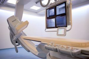
Sheep models can incorporate the safety and efficacy demands required for studies on orthopedic devices and implants, including the use of arthroscopy for more precise observation. Long bones such as the Tibia are superficial, therefore surgical access to them is easy.
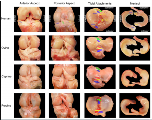
The knee: The width of the sheep medial-lateral tibial plateau is the largest of the animal models and therefore most similar to humans in size. The size and proportion of the human medial meniscus is most similar to the sheep and goat specimens. The picture below demonstrates the similarity between the human and sheep knee.
- In the picture below, the compatibility between the human and ovine knee and stifle joints are evident. For the tibial attachments, the following have been marked on both for additional clarity and comparison: ACL (anterior cruciate ligament), PCL (posterior cruciate ligament), AMM (anterior horn, medial meniscus), ALM (anterior horn, lateral meniscus), PMM (posterior horn, medial meniscus) and PLM (posterior horn, lateral meniscus).
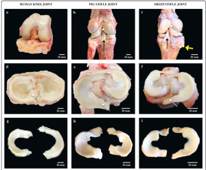
The similarity between the human knee joint and the sheep model stifle joint may not be easily understood. For further clarity, the view below gives a visual comparative of the knee joint and the stifle joint.

Sheep models have similar body weight and bone size as humans. They also present a similar bone in-growth pattern into porous scaffolds and both species present with comparable mineral compositions. This makes them advantageous for a wide range of preclinical studies and investigations.
Sheep models have been widely used in investigations involving:
- Orthopedic diseases (Intervertebral disc disease or IVDD, Degenerative disk disease, Spondylolisthesis, Spinal stenosis, Scoliosis, Fractured vertebra,
- Infection, Herniated disc, Tumor)
- Orthopedic implants and prostheses
- Fractures
- Osteoporosis
- Bone-lengthening
- Osteoarthritis
- Osseointegration of dental implants
- Menisci
- Intra Arterial injections (safety/efficacy)
- Osteoarthritis
- ACL (Anterior Cruciate Ligament)
- Spinal fusion
It is interesting to note that while bone healing of many animal models is faster than in humans, sheep models are considered to have a bone healing rate similar to humans. In a review article based on articles (1990 to 2016) found in PubMed, SciELO, LILACS, MEDLINE, and Cochrane, sighted that preclinical studies with sheep models were done without complications, allowing hypotheses to be both evaluated and mechanisms explained. Furthermore, the researchers concluded that sheep models are ‘excellent for evaluation of biomaterial for bone regenerations and dental implant osseointegration.’
Sheep models can incorporate the safety and efficacy demands required for studies on orthopedic devices and implants, including the use of arthroscopy for more precise observation.
Sartoretto, Suelen Cristina, et al. “Sheep as an experimental model for biomaterial implant evaluation.” Acta ortopedica brasileira 24 (2016): 262-266.