Introduction—Symptomatic mitral regurgitation (MR) is associated with high morbidity and mortality which can be
ameliorated by surgical valve replacement or repair. Despite this, many patients are excluded from surgery. Transcatheter mitral repair and replacement has emerged as a promising
therapeutic option for the treatment of severe MR. The majority of these procedures rely on high definition three
dimensional (3D) transesophageal echocardiographic (TEE) real time imaging. The sheep model is the model of choice for
preclinical studies of transcatheter mitral valve (MV) replacement and repair. However due to the thoracic conformation TEE studies in those animals are suboptimal.
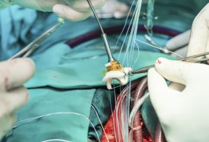
Methods and Results—In this study we tested and compared the feasibility of conventional TEE and newly described transpericardial 3D echocardiography (TPE) for assessment
of the MV as a tool to plan and possibly guide transcatheter/transapical procedures in the sheep model. The conventional TEE was challenging and produced incomplete
data in all of the animals. In contrast, the TPE method clearly depicted the left atria and ventricle, MV apparatus, LVOT, aortic valve and proximal ascending aorta facilitating high resolution 3D rendering in all of the animals. Conclusion—The value of TEE in the sheep model is limited and could not be reliable for complete assessment of the LV and mitral valve apparatus. Whereas, TPE produce reliable and similar imaging to TEE in the human patients which could allow animal testing of devices designed to be delivered in humans.